Services
Lameness Examinations
We carry our lameness examinations either at the clinic or owners’ property. A lameness work-up is usually made up of the following steps:
- Evaluation of the horse while standing and at walk, trot and canter if necessary.
- Flexion tests are often a useful part of lameness exam. These can sometimes temporarily exacerbate lameness and therefore help us to localise the source of pain.
- Perineural or intra-articular analgesia (nerve and joint blocks) involve injecting local anaesthetic either around nerves or directly into joints. By desensitizing particular areas and evaluating the difference in lameness the pain can be localized further.
- Imaging of the suspicious regions is then usually performed to look for abnormalities in the bones or soft tissue structures.
Our lameness exams generally take 1 to 2 hours. Lameness may be caused by several factors which require careful evaluation. At the end of the investigation, the consulting veterinarian will provide you with a diagnosis of your horse and recommend treatment options.
More complex lameness cases may require an in-depth investigation by our specialist clinicians, using advanced imaging techniques, such as CT or scintigraphy (bone scanning).
Lameness Assessments using the Equinosis Q
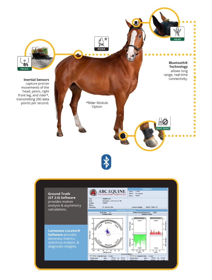
The Equinosis Q Lameness Locator is a real-time, handheld, field-based system that enables us to objectively measure lameness in horses with non-invasive inertial sensors. We have added this invaluable tool to our repertoire to help diagnose and manage ongoing lameness issues.
The equipment is made up of three sensors and a computer. One is placed on the head, one on the right pastern and one over the sacro-iliac joint. Each of these sensors plays an important role in the workings of the equipment. The head monitor measures the vertical movement of the head over time as we trot your horse up and down. The right pastern tells the equipment to know which stride is happening. As the right fore leg leaves the ground this notifies the machine that the left fore leg will be weight bearing. The pelvic sensors measure the vertical movement of the pelvis in time.
The correct placement of each of the sensors allows the computer to know what is happening when and where. In real time this data is relayed to the computer as we watch your horse trot either in a straight line or on the lunge or even under saddle.
Will it help me to see my horse’s lameness?
Yes! Gone are the days of quietly nodding in agreement with your vet, while you are unable to see what they are seeing! This assessment charts and plots lameness issues, so you can understand your horse’s issue/s better than ever. For any movement evaluation, the Equinosis Q is an excellent adjunctive device to diagnosis.
Call 9479 1800 to book your appointment!


View Our
Pricing Guide

View Our
Services Brochure


